Success Story

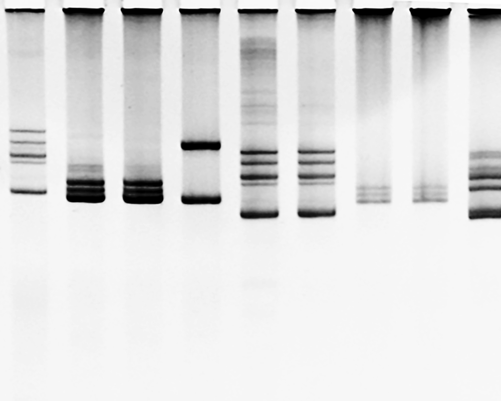
CCD imaging for Western Blot detection
April 19, 2021Make the transition from the traditional X-ray film developing to CCD imaging for Western Blot detection with Syngene chemiluminescence gel documentation systems.
Developing the blot on an X-ray film is the conventional way of detecting protein signals. It involves handling and developing the film in a dark room. The technique of film developing is less popular recently; mainly due to short-linearity of data for quantification.
CCD imagers, on the other hand, converts the chemical signals to digital images. Once the membrane is taken out of the developing solution, it is placed directly on the imaging platform of the CCD imager. The subsequent procedures are automated – with the hit of the camera button, the imager will take multiple different exposures of the blot and save all images electronically. Accurate quantification is achieved by densitometry. When appropriate standards are used, molecular weight analysis is made possible.
CCD imagers are much more convenient and accurate. Syngene's new generation of gel documentation systems promises high quantum efficiencies and superior sensitivity. Users are able to effortlessly capture their chemiluminescence and multiplex images without much "guessing".
NOTE TO EDITORSDr Yap Lai Lai
Department of Biochemistry, Yong Loo Lin School of Medicine,
National University of Singapore“ Our users are able to gain more knowledge on the use of the instruments and the analysis software which help significantly in our research. Now that we are introduced to this method, we aim to fully transition from X-ray film to scientific CCD cameras by end of the year”
You May Also Like
 16 April 2021
InAlyzer - World’s first 108-µm body composition analyzer
Use of high-resolution dual-energy X-ray Absorptiometry (DXA) for bone mineral density and body composition analysis
learn more